Overview of the Brain's Anatomy
NEURO
Injuries to the brain can be life-threatening. Normally the skull protects the brain from damage through its hard unyieldingness; the skull is one of the least deformable structures found in nature with it needing the force of about 1 ton to reduce the diameter of the skull by 1 cm.
CRANIAL NERVES
Your cranial nerves are pairs of nerves that connect your brain to different parts of your head, neck, and trunk. There are 12 of them, each named for their function or structure.
Each nerve also has a corresponding Roman numeral between I and XII. This is based on their location from front to back. For example, your olfactory nerve is closest to the front of your head, so it’s designated as I.
Their functions are usually categorized as being either sensory or motor. Sensory nerves are involved with your senses, such as smell, hearing, and touch. Motor nerves control the movement and function of muscles or glands.
I. Olfactory nerve
The olfactory nerve transmits sensory information to your brain regarding smells that you encounter.
II. Optic nerve
The optic nerve is the sensory nerve that involves vision.
III. Oculomotor nerve
The oculomotor nerve has two different motor functions: muscle function and pupil response.
Muscle function. Your oculomotor nerve provides motor function to four of the six muscles around your eyes. These muscles help your eyes move and focus on objects.
Pupil response. It also helps to control the size of your pupil as it responds to light.
This nerve originates in the front part of your midbrain, which is a part of your brainstem. It moves forward from that area until it reaches the area of your eye sockets.
IV. Trochlear nerve
The trochlear nerve controls your superior oblique muscle. This is the muscle that’s responsible for downward, outward, and inward eye movements.
V. Trigeminal nerve
The trigeminal nerve is the largest of your cranial nerves and has both sensory and motor functions.
The trigeminal nerve has three divisions, which are:
Ophthalmic. The ophthalmic division sends sensory information from the upper part of your face, including your forehead, scalp, and upper eyelids.
Maxillary. This division communicates sensory information from the middle part of your face, including your cheeks, upper lip, and nasal cavity.
Mandibular. The mandibular division has both a sensory and a motor function. It sends sensory information from your ears, lower lip, and chin. It also controls the movement of muscles within your jaw and ear.
VI. Abducens nerve
The abducens nerve controls another muscle that’s associated with eye movement, called the lateral rectus muscle. This muscle is involved in outward eye movement. For example, you would use it to look to the side.
VII. Facial nerve
The facial nerve provides both sensory and motor functions, including:
Moving muscles used for facial expressions as well as some muscles in your jaw
Providing a sense of taste for most of your tongue
Supplying glands in your head or neck area, such as salivary glands and tear-producing glands
Communicating sensations from the outer parts of your ear
VIII. Vestibulocochlear nerve
Your vestibulocochlear nerve has sensory functions involving hearing and balance. It consists of two parts, the cochlear portion and the vestibular portion:
• Cochlear portion. Specialized cells within your ear detect vibrations from sound based on the sound’s loudness and pitch. This generates nerve impulses that are transmitted to the cochlear nerve.
• Vestibular portion. Another set of special cells in this portion can track both linear and rotational movements of your head. This information is transmitted to the vestibular nerve and used to adjust your balance and equilibrium.
IX. Glossopharyngeal nerve
The glossopharyngeal nerve has both motor and sensory functions, including:
• Sending sensory information from your sinuses, the back of your throat, parts of your inner ear, and the back part of your tongue
• Providing a sense of taste for the back part of your tongue
• Stimulating the voluntary movement of a muscle in the back of your throat called the stylopharyngeus
X. Vagus nerve
The vagus nerve is a very diverse nerve. It has both sensory and motor functions, including:
• Communicating sensation information from your ear canal and parts of your throat
• Sending sensory information from organs in your chest and trunk, such as your heart and intestines
• Allowing motor control of muscles in your throat
• Stimulating the muscles of organs in your chest and trunk, including those that move food through your digestive tract (peristalsis)
• Providing a sense of taste near the root of your tongue
Out of all of the cranial nerves, the vagus nerve has the longest pathway. It extends from your head all the way into your abdomen.
XI. Accessory nerve
Your accessory nerve is a motor nerve that controls the muscles in your neck. These muscles allow you to rotate, flex, and extend your neck and shoulders.
It’s divided into two parts: spinal and cranial. The spinal portion originates in the upper part of your spinal cord. The cranial part starts in your medulla oblongata.
These parts meet briefly before the spinal part of the nerve moves to supply the muscles of your neck while the cranial part follows the vagus nerve.
XII. Hypoglossal nerve
Your hypoglossal nerve is the 12th cranial nerve which is responsible for the movement of most of the muscles in your tongue. It starts in the medulla oblongata and moves down into the jaw, where it reaches the tongue.
The brain is the most complex organ in the human body. It produces our every thought, action, memory, feeling, and experience of the world. This jelly-like mass of tissue, weighing in at around 1.4 kilograms in an adult, contains a staggering one hundred billion nerve cells, or neurons. The brain and spinal cord together form the central nervous system (CNS).
The complexity of the connectivity between these cells is mind-boggling. Each neuron can make contact with thousands or even tens of thousands of others, via tiny structures called synapses. Our brains form a million new connections for every second of our lives. The pattern and strength of the connections are constantly changing and no two brains are alike.
The neurons in our brains communicate in a variety of ways. Signals pass between them by the release of neurotransmitter and neuromodulator chemicals, such as glutamate, dopamine, acetylcholine, noradrenalin, serotonin, and endorphins.
Some neurochemicals work in the synapse, passing specific messages from release sites to collection sites, called receptors. Others also spread their influence more widely, like a radio signal, making whole brain regions more or less sensitive.
These neurochemicals are so important that deficiencies in them are linked to certain diseases. For example, a loss of dopamine in the basal ganglia, which controls movements, leads to Parkinson’s disease. It can also increase susceptibility to addiction because it mediates our sensations of reward and pleasure.
Similarly, a deficiency in serotonin, used by regions involved in emotion, can be linked to depression or mood disorders, and the loss of acetylcholine in the cerebral cortex is characteristic of Alzheimer’s disease.
The brain is made up of multiple parts, and each part of the brain is responsible for different body functions. Therefore, brain tumor symptoms depend a great deal on where the tumor is located.
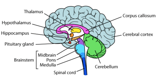
Major parts of the brain: There are three major parts of the brain.
Cerebrum: uses information from senses to tell our body how to respond. It controls reading, thinking, learning, movement, speech, vision, personality, and emotions.
Cerebellum: controls balance for standing, walking, and other motion.
Brain stem: connects the brain with the spinal cord and controls basic body functions such as breathing, sleeping, body temperature, and blood pressure.

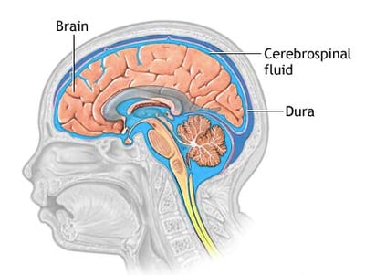
It is protected by:
• The skull bones
• 3 fine layers of tissue (meninges-dura, arachnoid, and pia)
• Cerebrospinal fluid (Watery fluid) flowing through spaces between the meninges and through spaces (ventricles) within the brain.

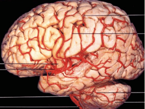

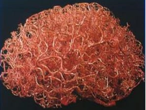

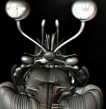
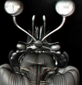
Brain Vasulature
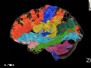
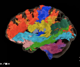
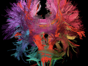

Brain Fibers
SKULL
The skull is a bone structure that forms the head in vertebrates. It supports the structures of the face and provides a protective cavity for the brain. The skull is composed of two parts: the cranium and the mandible
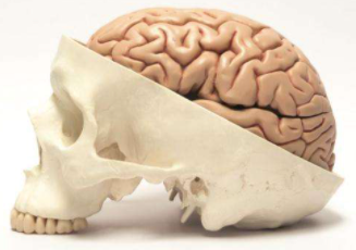

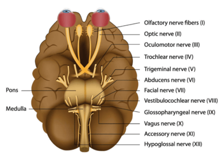
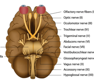
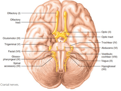

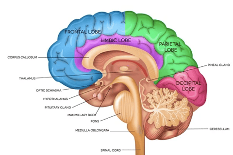
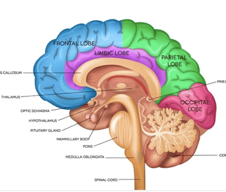
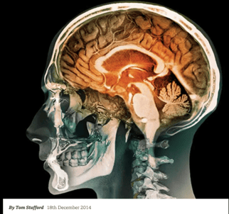
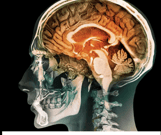
Image Source: Neuroanatomy by Martin C Hirsch & Thomas Kramer
© Copyright2024 - Dr Ashok Hande - All rights reserved
Clinic Details
Timings: 11 AM - 8 PM
Fortis Hospital
Contact Details for Emergency(24 Hrs)